

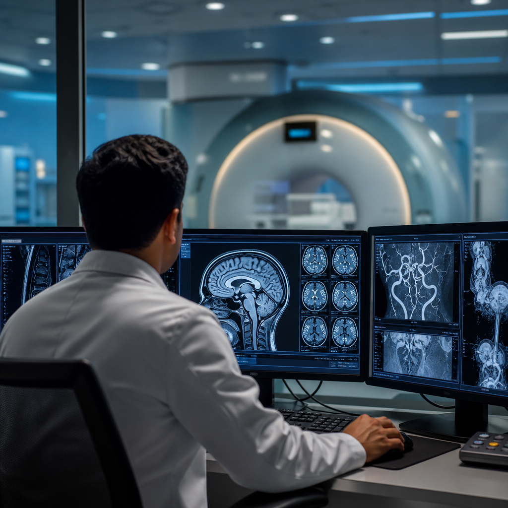
The human nervous system is among the most intricate structures in existence, and diagnosing its disorders demands an equally sophisticated approach. Over the past two decades, neuroradiology has evolved from a supporting discipline into one of the most critical pillars of neurological care. From detecting early-stage tumors to mapping the precise anatomy of a stroke, the imaging technologies used today give clinicians an unprecedented window into the brain and spinal cord. As patient expectations rise and conditions become more complex, the role of specialized imaging centers has never been more consequential.
What Neuroradiology Actually Involves
Neuroradiology is a subspecialty of radiology focused exclusively on diagnosing and, in some cases, treating conditions of the central and peripheral nervous system. This includes the brain, spinal cord, spine, head, and neck. Unlike general radiology, neuroradiology requires deep familiarity with neuroanatomy, pathology, and the nuanced ways disease manifests across imaging modalities.
The tools of the trade include magnetic resonance imaging (MRI), computed tomography (CT), positron emission tomography (PET), and digital subtraction angiography (DSA). Each modality serves a different diagnostic purpose. MRI, for instance, offers exceptional soft tissue contrast and is the gold standard for evaluating brain tumors, multiple sclerosis, and white matter disease. CT, on the other hand, is faster and better suited for acute trauma or hemorrhage detection. Functional MRI (fMRI) and diffusion tensor imaging (DTI) have added further layers of capability, allowing clinicians to map brain activity and white matter tracts with remarkable precision.
Interventional Neuroradiology: Beyond Diagnosis
One of the most significant developments in the field is the rise of interventional neuroradiology, sometimes called endovascular neurosurgery. Rather than simply identifying a problem, interventional neuroradiologists can treat it directly through minimally invasive catheter-based procedures. Conditions such as cerebral aneurysms, arteriovenous malformations (AVMs), and acute ischemic stroke can now be addressed through the blood vessels themselves, often without a single surgical incision. This approach dramatically reduces recovery times and complication rates compared to open surgery, making it a preferred option for many patients.
The Growing Demand for Specialized Neurological Imaging
Neurological conditions are on the rise globally. Alzheimer’s disease, Parkinson’s disease, epilepsy, brain tumors, and spinal disorders collectively affect hundreds of millions of people worldwide. As populations age and awareness of neurological health increases, demand for high-quality diagnostic imaging has surged correspondingly. Patients and referring physicians alike are seeking centers that combine cutting-edge technology with subspecialty expertise — not just general imaging departments that handle neurological cases as one of many categories.
This shift in demand mirrors a broader trend in healthcare: the move toward specialization. Just as patients with complex cardiac conditions seek out dedicated heart centers, those with neurological concerns increasingly want access to teams whose entire focus is the nervous system. The quality of interpretation matters enormously in neuroradiology. A subtle finding on an MRI — a small area of restricted diffusion, a barely perceptible mass effect — can be the difference between a timely diagnosis and a missed one.
Digital Fatigue and Its Neurological Consequences
Modern lifestyles are placing unprecedented strain on the nervous system and sensory organs alike. Prolonged screen exposure, irregular sleep patterns, and chronic stress are contributing to a range of neurological complaints, from persistent headaches and visual disturbances to cognitive fog and anxiety disorders. It is worth noting that screen fatigue is driving a notable spike in eye-related health inquiries, reflecting how interconnected our visual and neurological systems truly are. Neuroradiologists are increasingly being called upon to evaluate patients whose symptoms originate at this intersection — where lifestyle factors meet measurable neurological change.
Neuroradiology’s Critical Role in Oncology
Brain and spinal tumors represent some of the most challenging diagnoses in medicine. Neuroradiology plays an indispensable role not only in initial detection but throughout the entire treatment journey. Before surgery, detailed imaging helps neurosurgeons plan their approach, identifying eloquent cortex areas that must be preserved. During treatment, follow-up MRIs track tumor response and detect recurrence. After therapy, imaging distinguishes between true progression and treatment-related changes such as pseudoprogression — a distinction that has profound implications for patient management.
For patients preparing for brain or spinal surgery, understanding what lies ahead is essential. Comprehensive resources on preparing for cancer surgery can help patients and families navigate the process with greater confidence and clarity. Neuroradiologists work closely with oncologists and neurosurgeons to ensure that imaging findings are communicated clearly and translated into actionable treatment plans.
Artificial Intelligence and the Future of Neuroimaging
Artificial intelligence is beginning to transform neuroradiology in meaningful ways. Machine learning algorithms trained on vast datasets of neuroimaging studies can now assist radiologists in detecting anomalies, quantifying lesion burden, and even predicting disease progression. AI tools are particularly promising in conditions like multiple sclerosis, where tracking lesion load over time is critical, and in stroke imaging, where speed of diagnosis directly impacts outcomes. However, AI in neuroradiology is best understood as an augmentation tool rather than a replacement for expert human judgment. The subtleties of neuroimaging interpretation — the clinical context, the patient history, the nuanced pattern recognition built over years of subspecialty training — remain firmly in the domain of the specialist.
Cleveland Clinic Abu Dhabi: A Center of Neuroimaging Excellence
For patients in the UAE and the broader region seeking world-class neurological imaging, Cleveland Clinic Abu Dhabi stands as a benchmark institution. The neuroradiology department at Cleveland Clinic Abu Dhabi brings together fellowship-trained subspecialists, state-of-the-art imaging technology, and a multidisciplinary care model that ensures findings are integrated seamlessly into each patient’s overall treatment plan. Whether the clinical question involves a complex brain tumor, a vascular malformation, a degenerative spine condition, or an acute neurological emergency, the team is equipped to provide precise, timely, and clinically meaningful answers. The department’s commitment to diagnostic accuracy and patient-centered care reflects the broader ethos of an institution that has consistently set the standard for specialty medicine in the region.
Conclusion: Precision Imaging as the Foundation of Neurological Care
Neuroradiology has moved far beyond its origins as a purely interpretive discipline. Today, it sits at the intersection of diagnosis, treatment planning, intervention, and ongoing disease monitoring. As neurological conditions grow more prevalent and treatment options more sophisticated, the quality of imaging and interpretation becomes ever more consequential. Patients deserve access to specialists who understand not just how to operate an MRI scanner, but how to extract every clinically relevant detail from the images it produces — and how to communicate those findings in ways that drive better outcomes. In a field where precision is everything, the choice of where to seek neuroimaging care is a decision that genuinely matters.
